


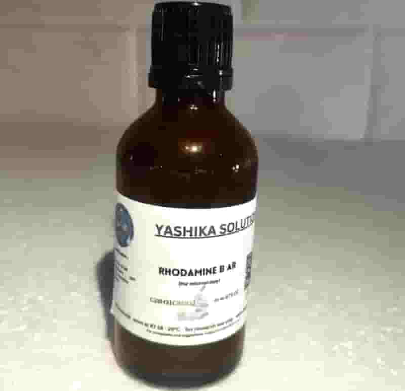

Rhodamine B AR for Microscopy
Customize
Product Details
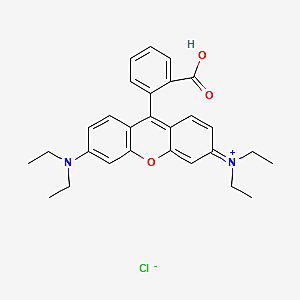
Rhodamine B is a fluorescent dye commonly used in microscopy for various applications due to its bright and stable fluorescence properties. Some of its uses in microscopy include:
Cell Staining: Rhodamine B can be conjugated with antibodies or other molecular probes to specifically label cellular structures, such as nuclei, cytoskeleton, or organelles. This allows researchers to visualize and study the morphology, dynamics, and interactions of different cellular components under a microscope.
Live Cell Imaging: Rhodamine B is often used as a vital dye to label living cells. Its fluorescence can be excited using a suitable light source, allowing real-time imaging of cellular processes, such as cell migration, division, or signal transduction, without compromising cell viability.
Fluorescence Microscopy: Rhodamine B is compatible with various fluorescence microscopy techniques, including confocal microscopy, widefield fluorescence microscopy, and super-resolution microscopy. Its intense fluorescence emission and resistance to photobleaching make it suitable for imaging highly dynamic cellular processes with high spatial resolution.
Tissue Labeling: In histology and pathology, Rhodamine B can be used to label tissues and biopsy samples for examination under a microscope. It allows visualization of tissue architecture, cell types, and pathological changes, aiding in the diagnosis of diseases and assessment of tissue morphology.
Intracellular pH Measurement: Rhodamine B is sensitive to changes in pH, and its fluorescence properties can be used to monitor intracellular pH dynamics in living cells. By loading cells with Rhodamine B and measuring changes in fluorescence intensity, researchers can investigate pH-dependent cellular processes and signaling pathways.
Overall, Rhodamine B is a versatile fluorescent dye widely used in microscopy for labeling cells, tissues, and subcellular structures, as well as for dynamic imaging and functional studies in biological research. Its bright and stable fluorescence, compatibility with various imaging techniques, and pH sensitivity make it an invaluable tool for studying complex biological systems at the microscopic level.