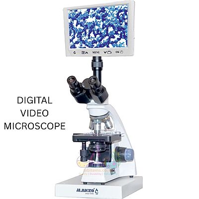
DIGITAL VIDEO MICROSCOPE – DVM-02 Plus
The DVM-02 Plus Digital Video Microscope is an advanced research and teaching microscope integrated with a 9.7” IPS LCD digital display and Android-based software platform. Designed for real-time observation, measurement, analysis, and documentation, it combines precision optical microscopy with powerful digital imaging and connectivity features.
With trinocular viewing, high-resolution CMOS camera, HDMI output, SD card storage, and built-in analysis software, the DVM-02 Plus is ideal for medical colleges, research laboratories, pathology labs, quality control, and training institutes.
Key Features & Benefits
| Feature | Benefit |
|---|
| 9.7” IPS LCD Digital Display | Large, clear screen for comfortable real-time viewing |
| Android-Based Operating System | Smooth operation with built-in analysis software |
| High-Resolution CMOS Camera | Accurate image capture for documentation and reporting |
| Trinocular Viewing Head | Supports simultaneous eyepiece and digital observation |
| Built-in Measurement & Scale Software | Enables precise measurement and particle analysis |
| HDMI Output & SD Card Support | Easy data sharing, storage, and external display |
| Co-Axial Focusing System | Stable and precise focusing for research-grade work |
| Inbuilt Battery Backup | Ensures uninterrupted operation |
Optical Specifications
| Parameter | Specification |
|---|
| Viewing Head | Trinocular head, 30° inclined, 360° rotatable |
| Interpupillary Distance | 54 – 75 mm |
| Diopter Adjustment | ±5 on left tube |
| Eyepiece | Wide Field WF 10× / 18 mm |
| Objectives | Plan Achromatic 4×, 10×, 40×, 100× (oil) |
| Nosepiece | Quadruple revolving nosepiece |
| Condenser | Abbe Condenser NA 1.25 with iris diaphragm & filter holder |
Mechanical & Focusing System
| Parameter | Specification |
|---|
| Focusing | Co-axial coarse and fine focusing |
| Fine Focus Precision | 0.002 mm |
| Coarse Stroke | 36 mm with adjustable rack stop |
| Friction Control | Adjustable coarse focus friction |
| Stage Type | Double layer mechanical stage |
| Stage Size | 110 mm × 125 mm |
| Movement Range | 30 mm × 75 mm |
Illumination & Power System
| Parameter | Specification |
|---|
| Light Source | 3W LED illumination |
| Brightness Control | Adjustable intensity |
| Power Adapter | External 110–240V AC adapter |
| Battery Backup | Inbuilt battery backup facility |
| Power Input | DC 5V / 230V AC |
Camera Specifications
| Parameter | Specification |
|---|
| Image Sensor | 1/2.5” CMOS, 5.0 Megapixel |
| Valid Pixels | 2592 × 1944 |
| Pixel Size | 2.2 μm × 2.2 μm |
| Dynamic Range | 66.5 dB |
| Resolution & Frame Rate | 1280×720 @15 fps, 640×480 @30 fps |
| Signal-to-Noise Ratio | 40.5 dB |
| Sensitivity | 0.53 V/lux-sec (550 nm) |
Tablet / Display Specifications
| Parameter | Specification |
|---|
| Display Size | 9.7” IPS LCD |
| Resolution | 1024 × 768 |
| RAM | 1 GB DDR3 |
| Internal Storage | 2 GB |
| External Storage | SD card support up to 32 GB |
| Operating System | Android 4.2.2 |
| Connectivity | HDMI output, SD card, Bluetooth |
| Software | Built-in microscopy & measurement software |
Applications
| Field | Usage |
|---|
| Research Laboratories | Digital imaging, analysis & documentation |
| Medical Colleges | Teaching pathology, histology & microbiology |
| Diagnostic Labs | Routine examination and reporting |
| Training Institutes | Group demonstrations and learning |
| Quality Control | Inspection, measurement & image recording |
Packaging & Delivery
| Item | Details |
|---|
| Inner Packing | Thermocol / Styrofoam |
| Outer Packing | Corrugated cardboard box |
| Shipping | Secure packaging suitable for transport |